By Dr. Prateek Porwal, ENT Surgeon & Vertigo Specialist | PRIME ENT Center, Hardoi UP
Last Updated: February 2026 | VAI Budapest 2025 Award Recipient
The most critical part of treating vertigo is getting the diagnosis right. I can’t tell you how many patients arrive at my clinic after spending thousands of rupees on unnecessary imaging because their previous doctor didn’t perform a proper clinical evaluation. A good history and physical exam solve 90% of vertigo cases without needing advanced testing. This article walks you through exactly how I diagnose vertigo at PRIME ENT Center—the systematic approach that leads to accurate diagnosis, most of the time in a single visit.
The Diagnostic Hierarchy: Why Order Matters
Here’s what many doctors get wrong: they order tests first and ask questions later. The correct approach is history first, physical exam second, tests only when needed. This hierarchy exists for a reason—it’s the most efficient and cost-effective path to diagnosis. In India, where patients often come with limited resources, skipping the expensive parts when not needed is important.
Step 1: Taking a Detailed History – The Foundation of Diagnosis
Image Source: Canva
I spend 10-15 minutes taking history. This isn’t idle chat—specific questions narrow the diagnosis dramatically. Here are the five questions that crack 80% of vertigo diagnoses:
Question 1: Onset – How Did This Start?
Sudden onset (minutes to hours) suggests vestibular neuritis, BPPV, labyrinthitis, or stroke. Gradual onset (days to weeks) suggests migraine, tumor, or central cause. The key detail: “I was fine when I woke up, then suddenly the room started spinning” is BPPV or vestibular neuritis. “I’ve had this for 3 weeks and it’s getting slowly worse” is different—needs imaging.
Question 2: Duration – How Long Does Each Episode Last?
Seconds to 1 minute: Classic BPPV. Patient gets vertigo when turning over in bed or rolling their head. Minutes to hours: Vestibular migraine. Hours to days: Vestibular neuritis or labyrinthitis. Constant for weeks: Central cause, PPPD, or chronic vestibular dysfunction. Duration is one of the most reliable diagnostic clues.
Question 3: Recurrence Pattern – Is This One Episode or Multiple?
Single episode that’s resolving: Probably vestibular neuritis. Recurring episodes: BPPV, Meniere’s disease, or vestibular migraine. The timeline: “It happens twice a month” vs. “It happens once a year” changes the diagnosis. For BPPV, I see patients who have one episode, recover, then have another 3 months later. For Meniere’s, attacks are more regular initially but eventually spread further apart.
Question 4: Trigger – What Sets It Off?
Specific head position triggers: BPPV. “It happens when I turn over in bed” or “When I look up” is almost diagnostic. Stress and hormones: Vestibular migraine. Pressure changes or loud noise: Superior canal dehiscence or perilymph fistula. No clear trigger: Vestibular neuritis or central cause. The trigger question is gold—it directly points to mechanism.
Question 5: Vertigo vs. Lightheadedness – What Exactly Do You Feel?
“The room spins”: True vertigo, peripheral cause. “I feel faint/lightheaded”: Non-vertigo dizziness, consider cardiovascular, metabolic, or central. This distinction is critical because treatments differ completely. A patient who says “I feel like I’m about to faint” doesn’t need vestibular rehab; they need their blood pressure checked.
Associated Symptoms – The Secondary Clues
Hearing loss or tinnitus? Suggests inner ear involvement—BPPV doesn’t cause hearing loss, but Meniere’s disease and labyrinthitis do. Headache? Suggests vestibular migraine. Weakness, numbness, or speech problems? Red flag for stroke—needs imaging urgently. Fever or recent infection? Suggests viral labyrinthitis. Recent head injury? Trauma-related, possibly perilymph fistula.
Medication History – Often Overlooked
New medications can cause dizziness. Ototoxic drugs (certain antibiotics like gentamicin or aminoglycosides, some cancer drugs) can damage hearing and balance. I always ask: “Did your vertigo start after starting any new medication?” If yes, medication may be the culprit. Some patients have been dizzy for months because their doctor didn’t connect it to a medication started around that time.
Step 2: Physical Examination – Where Diagnosis Happens
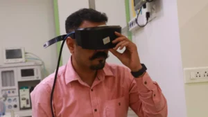
Image Source: Canva
Physical exam is where diagnosis happens. Here’s what I do:
Bedside Positional Tests:
- Dix-Hallpike Test – I rapidly tip your head back over the edge of the examination table. If you get vertigo and I see characteristic nystagmus (eye jerking), BPPV is confirmed. This single test diagnoses 80% of my vertigo patients. Positive test = BPPV treatment today, no imaging needed
- Supine Roll Test – For BPPV in the horizontal canal. Patient lies supine, head hanging off table, I rotate head to one side. Different canal involvement = different positioning maneuver needed
- Romberg Test – Standing with eyes closed to assess balance and proprioception. Falling when eyes closed = vestibular dysfunction
- Unterberger (Fukuda) Stepping Test – Patient marches in place with eyes closed. Turning toward one side suggests that side’s vestibular weakness
- Gait Assessment – How you walk tells me a lot about balance function. Wide-based gait, veering to one side, inability to walk straight line all suggest vestibular involvement
Eye Movement Examination – The HINTS Exam:
This is critical. I perform three specific tests to differentiate between peripheral and central vertigo:
- H = Head Impulse Test (Doll’s Head Maneuver) – I move your head quickly while you keep eyes on a target. Normal response: Eyes stay on target, head moves. Abnormal response: Eyes lag behind head movement, suggesting vestibular weakness
- I = Nystagmus Assessment – I observe eye movements. Peripheral vertigo nystagmus: Unidirectional, goes away with fixation on a target. Central vertigo nystagmus: Can be in any direction, persists with fixation
- T = Test of Skew – I cover one eye at a time looking for vertical misalignment. Skew deviation (one eye higher than the other) is a red flag for stroke
Why HINTS matters: This exam is more accurate than MRI in the first 24 hours of a stroke affecting the cerebellum. Stroke can present as vertigo, and missing it is dangerous. If HINTS is abnormal, that patient gets urgent MRI and neurology evaluation.
Additional Eye Movement Tests:
- Smooth Pursuit – Can you smoothly follow a moving target? Abnormality suggests central pathology
- Saccades – Can you make quick eye movements between targets? Impaired saccades suggest brainstem or cerebellar disease
- Caloric Response – Does your eye respond normally to vestibular stimulation? I don’t do formal caloric testing in clinic, but bedside assessment matters
Neurological Assessment:
I check for weakness, numbness, coordination abnormalities, or other neurological signs that would suggest central cause requiring urgent MRI. If neuro exam is abnormal in any way with vertigo, imaging is needed.
Step 3: Specialized Testing (When Necessary)
Image Source: Canva
Most BPPV diagnoses need no testing beyond clinical examination. But some cases require:
Vestibular Function Tests:
- VNG (Videonystagmography) – Objective measurement of eye movements during vestibular testing using infrared eye tracking. I have this at PRIME ENT Center. Useful for: detecting subtle nystagmus, objective documentation of vestibular response, VNG-supported Dix-Hallpike testing for medico-legal cases
- Caloric Testing – Warm and cold water (or air) in the ear canal to test vestibular response. Compares left and right vestibular function. Particularly useful for detecting unilateral vestibular weakness. Less commonly used now but valuable in specific cases
- Rotatory Chair Testing – For complex cases. Patient sits in chair that rotates while eye movements are tracked. Provides detailed information about vestibular system function across different frequencies of head movement. Rarely needed in primary care but useful for research and complex cases
- Dynamic Posturography – Patient stands on moving platform. The platform and visual surroundings move, testing how they maintain balance. Useful for assessing fall risk and functional recovery in rehabilitation
Hearing Tests:
- Audiometry – Formal hearing test. Important for Meniere’s disease diagnosis (shows low-frequency hearing loss initially), labyrinthitis (can show sensorineural hearing loss), vestibular neuritis (hearing usually normal, so abnormal audiometry suggests different diagnosis)
- Tympanometry – Assesses middle ear function. Helps differentiate between conductive and sensorineural hearing loss
- Acoustic Reflex Testing – Tests stapedial muscle response. Abnormality suggests facial nerve involvement or acoustic neuroma
Imaging Studies – When and Why:
- MRI Brain and Internal Auditory Canal – For suspected central causes (stroke, tumor, MS), progressive symptoms, asymmetric hearing loss (rules out acoustic neuroma), chronic unexplained vertigo. Note: Most BPPV doesn’t need MRI. Ordering MRI for obvious BPPV is wasteful
- CT Temporal Bones – Faster than MRI if stroke suspected and MRI unavailable. Shows bone anatomy better—useful for superior canal dehiscence, perilymph fistula, or temporal bone fracture
- Common Error: Ordering imaging for every dizzy patient. In my experience, 50-60% of patients who see non-specialists get “routine” MRI when clinical exam clearly indicates BPPV. This wastes patient money and healthcare resources
Blood Tests:
- Usually not necessary for pure vertigo diagnosis
- Helpful if metabolic cause suspected (low glucose, anemia, thyroid dysfunction, vitamin deficiency)
- Blood pressure measurements (sitting and standing) assess orthostatic changes
- Consider if fever and vertigo suggest infection
My Diagnostic Algorithm – How I Actually Think Through Cases
Brief, position-triggered vertigo, classic nystagmus on Dix-Hallpike? BPPV. No imaging needed. Treatment today. Patient goes home with repositioning instructions, often improved immediately.
Sudden severe vertigo, days of duration, no hearing loss, normal HINTS exam? Vestibular neuritis. Maybe VNG to confirm. Rest, vestibular exercises, gradual activity increase.
Sudden severe vertigo WITH hearing loss and tinnitus? Labyrinthitis or Meniere’s disease. Audiometry helps confirm hearing loss pattern. May need imaging if first episode and concern for retrocochlear pathology.
Recurrent vertigo with hearing loss, tinnitus, aural fullness, and episodes lasting hours? Meniere’s disease. Audiometry and imaging (to rule out acoustic neuroma causing similar symptoms). Salt-restricted diet, diuretics, migraine prophylaxis.
Vertigo with migraine history, or episodes lasting minutes to hours without positional trigger? Vestibular migraine. VNG if any question about vestibular function. Migraine prevention is treatment.
Vertigo with ANY neuro symptoms (weakness, numbness, speech, gait abnormality, abnormal HINTS)? Central cause suspected. Need urgent MRI and neurology evaluation.
Chronic unsteadiness for months, constant symptoms, normal positional tests? PPPD (Persistent Postural-Perceptual Dizziness), bilateral vestibular hypofunction, or central cause. Requires VNG and possibly imaging to characterize.
Advanced Technique: The Bangalore Maneuver for Complex BPPV
For BPPV that doesn’t respond to standard Epley maneuver or has multiple canal involvement, I use the Bangalore Maneuver—an advanced repositioning technique that combines multiple sequences. This approach significantly improves success rates in complex BPPV cases where crystals are stubborn or patient anatomy is complex.
Diagnosis at PRIME ENT Center – The One-Visit Approach
My goal is diagnosis and initial treatment in one visit. Here’s what happens:
Visit 1 (typically 45 minutes): History, Dix-Hallpike test, HINTS exam, other bedside tests, diagnosis made. If BPPV, Epley or other repositioning maneuver done during same visit. Patient often improves within hours. If not BPPV, discuss treatment plan.
Follow-up as needed: Only if: diagnosis unclear after initial evaluation, patient not improving as expected, or complex case requiring VNG or imaging.
This approach saves patients time and money while improving outcomes through rapid diagnosis and treatment.
Frequently Asked Questions
Do I need an MRI for vertigo?
Not always. If clinical exam and history suggest BPPV, Meniere’s, or vestibular neuritis, MRI isn’t necessary. MRI is important for suspected central causes, chronic unexplained vertigo, progressive symptoms, or abnormal neurological exam.
Can a CT scan diagnose vertigo?
CT is better than MRI for showing bone detail but less useful for soft tissue. For most vertigo diagnosis, clinical exam is primary. Imaging is confirmatory, not diagnostic.
How accurate is the Dix-Hallpike test?
Very accurate in trained hands. Sensitivity is high for posterior canal BPPV. False negatives can occur if crystals haven’t migrated into the canals yet. False positives are rare if physician knows what positive nystagmus looks like.
What if tests are normal but I still have vertigo?
Normal tests don’t mean vertigo is imaginary. Some conditions (like PPPD or central sensitization) don’t show abnormalities on standard tests. Diagnosis may rely more on history and pattern recognition. Consider second opinion from vestibular specialist.
How long does diagnosis take?
A proper exam takes 30-45 minutes. BPPV diagnosis is often immediate during Dix-Hallpike. Other diagnoses may need observation over time or repeat testing to confirm.
Should I get a second opinion?
Yes, if diagnosis is unclear, treatment isn’t working, or you’re about to have surgery. Getting another specialist’s perspective is reasonable, especially from someone experienced in vestibular disorders.
What is the HINTS exam?
The HINTS exam (Head Impulse, Nystagmus, Test of Skew) is a bedside test that’s more accurate than MRI in the first hours of a cerebellar stroke. It helps differentiate peripheral vertigo from central causes.
Why is VNG testing useful?
VNG provides objective documentation of eye movements and vestibular response. It’s useful for detecting subtle abnormalities, confirming clinical findings, and tracking change over time with treatment.
Can vertigo be diagnosed without imaging?
Absolutely. Most BPPV, vestibular neuritis, and many other conditions are diagnosed clinically without imaging. Imaging is for specific indications, not routine screening.
What is the most common vertigo diagnosis?
BPPV (Benign Paroxysmal Positional Vertigo) is by far the most common, accounting for 40-50% of all vertigo cases. It’s easily diagnosed with Dix-Hallpike test and highly treatable with repositioning maneuvers.
Experiencing vertigo or chakkar? Get diagnosed — usually in one visit.
Dr. Prateek Porwal, ENT Surgeon & Vertigo Specialist at PRIME ENT Center, Hardoi UP — VAI Budapest 2025 International Award recipient. Most BPPV cases resolved in the same appointment.
Call/WhatsApp: 7393062200 | Chat on WhatsApp