This vertigo/”>vestibular rehabilitation evidence-based protocol article summarises the neurophysiological basis of vestibular compensation, patient selection criteria, disorder-specific exercise protocols, and outcome predictors for clinical practice — grounded in Level 1 evidence (Hillier & McDonnell, Cochrane 2011). It is intended as a reference for ENT surgeons, neurologists, and vestibular physiotherapists managing patients with chronic vertigo-statistics/”>dizziness and balance disorders.
This is a glimpse of Dr Prateek Porwal’s vestibular rehabilitation evidence-based protocol. For a detailed and specific VRT programme tailored to individual patient presentations, contact Dr Prateek Porwal.
For patients: see the patient-facing guide to vestibular rehabilitation exercises.
Overview: Vestibular Rehabilitation Evidence-Based Protocol and Clinical Framework
Effective vestibular rehabilitation begins with thorough patient assessment and structured counselling. The clinical consultation should address the patient’s primary concerns — particularly fall risk and mobility limitation — establish realistic outcome expectations, and document aggravating factors and relevant comorbidities including hypertension, diabetes, and anxiety disorders. Patient engagement and compliance are critical determinants of rehabilitation success; outcome data consistently show that adherence to prescribed exercise frequency is the strongest predictor of recovery rate.
Investigations to contextualise the rehabilitation programme include pure tone audiometry (PTA), video nystagmography (VNG), vestibular evoked myogenic potentials (VEMP), and auditory brainstem response (ABR/BERA) where indicated. These establish the laterality, severity, and nature of vestibular dysfunction and guide exercise selection and progression criteria.
Common Presenting Features in Vestibular Patients
The six cardinal symptoms presenting in vestibular clinic are: vertigo and dizziness; anxiety and depression; nausea and autonomic symptoms; imbalance and gait disturbance; reduced quality of life; and recurrent episodic attacks. Clinical reasoning should prioritise therapeutic time on symptoms 2 through 6. Psychological and functional factors — particularly anxiety-driven avoidance behaviour — significantly predict suboptimal outcomes and require concurrent management alongside physical rehabilitation.
Holistic Management Considerations
Rehabilitation planning should incorporate lifestyle and dietary factors with documented impact on vestibular function. Recommended baseline guidance includes adequate hydration (minimum 2 litres water daily), reduced caffeine and dietary salt, avoidance of alcohol, and stress reduction interventions. Tai chi and yoga have Level B evidence for balance improvement in vestibular populations and serve as useful adjuncts to formal VRT. Personalising the programme to the patient’s occupational and social context improves long-term compliance.
Neurophysiological Basis of the Vestibular Rehabilitation Evidence-Based Protocol
Central compensatory neuroplasticity — the cornerstone of any vestibular rehabilitation evidence-based protocol — involves VOR gain recalibration and cerebellar synaptic remodelling. Following unilateral peripheral lesion, the brainstem experiences tonic imbalance between ipsilesional and contralesional vestibular nuclei. Compensation proceeds via three mechanisms: static compensation (resolution of spontaneous nystagmus and postural asymmetry at rest), dynamic compensation (partial recovery of VOR gain during head movement), and substitution (use of smooth pursuit, cervico-ocular reflex, and somatosensory inputs to supplement deficient vestibular drive).
Adaptation exercises leverage Hebbian plasticity principles: repetitive mismatched visual-vestibular input drives error-signal-dependent synaptic remodelling in the flocculus and nodulus. The adaptation pathway is preferred when VOR gain deficit is the primary functional impairment, as quantified by video head impulse test (vHIT) or caloric asymmetry. Habituation protocols are appropriate when symptom provocation rather than VOR gain deficit is the dominant complaint, as in PPPD.
Clinical Assessment Tools
The following standardised outcome measures form the core of any vestibular rehabilitation evidence-based protocol, and should be applied at baseline and at four-weekly intervals:
- Dizziness Handicap Inventory (DHI) — 25-item self-report scale assessing functional, emotional, and physical impact of dizziness. Score >18 indicates moderate-severe handicap. Minimally clinically important difference (MCID): 18 points.
- Functional Gait Assessment (FGA) — 10-item observational gait tool sensitive to vestibular-related gait impairment. Score <22/30 predicts fall risk in vestibular populations.
- Modified Clinical Test of Sensory Integration for Balance (mCTSIB) — Four-condition standing test (firm/foam surface × eyes open/closed) quantifying sensory weighting. Essential for bilateral vestibulopathy assessment.
- Video Head Impulse Test (vHIT) — Objective VOR gain measurement per semicircular canal plane. Gain <0.8 with corrective saccades confirms canal-specific hypofunction.
- Caloric Test — Canal paresis >25% (Jongkees formula) indicates significant unilateral hypofunction; guides prognosis for compensation rate.
- Subjective Visual Vertical (SVV) — Deviation >2.5° indicates utricular dysfunction; useful in PPPD assessment when combined with moving visual background.
Patient Selection and Outcome Predictors
Factors associated with favourable vestibular rehabilitation evidence-based protocol outcomes include younger age, shorter symptom chronicity, unilateral rather than bilateral dysfunction, absence of central pathology on neuroimaging, and absence of significant anxiety or depression. Patients with higher pre-treatment VOR gain deficit and greater caloric asymmetry paradoxically achieve stronger objective gains from adaptation-based protocols, as the error signal driving cerebellar plasticity is larger. Patient anxiety and avoidance behaviour predict suboptimal outcomes; consider concurrent CBT referral in patients with elevated anxiety scores (GAD-7 ≥10) or DHI emotional subscale scores ≥36.
Negative predictors include: unstable Menière’s disease with frequent attacks, active central lesion (acoustic neuroma recurrence, demyelination, cerebellar stroke), uncontrolled cardiovascular disease limiting exercise tolerance, significant cervicogenic dizziness as the primary driver (requires concurrent cervical physiotherapy), and long-term vestibular suppressant use (vestibular suppressants attenuate the error signal necessary for adaptation and should be tapered prior to commencing VRT).
Evidence-Based Protocol: Dosage Parameters by Condition
| Condition | VRT Frequency | Expected Recovery Window | Evidence Level |
|---|---|---|---|
| Acute unilateral vestibular neuritis | 3–5 sessions/day, 10–15 min each | 6–8 weeks (functional); 3–6 months (VOR gain) | Level A (Cochrane 2011) |
| Uncompensated unilateral hypofunction | 3–4 sessions/day, 15–20 min each | 8–12 weeks | Level A |
| Bilateral vestibulopathy | 2–3 sessions/day, 20 min each; supervised initially | 12–24 weeks (partial); ongoing maintenance | Level B (Strupp et al. 2017) |
| PPPD (combined VRT + CBT) | Daily VRT + weekly CBT sessions | 12–16 weeks | Level B (Staab et al. 2017) |
| Post-acoustic neuroma surgery | 3–4 sessions/day commencing 2–4 weeks post-op | 12–26 weeks | Level B |
| Age-related balance decline (presbyvestibulia) | 2–3 sessions/day; supervised for fall-risk patients | 8–16 weeks; maintenance ongoing | Level B |
Unilateral Vestibular Loss — Vestibular Rehabilitation Evidence-Based Protocol
Acute Phase
Acute unilateral vestibular loss (vestibular neuritis, labyrinthitis) presents with severe nausea, rotary vertigo, spontaneous nystagmus with oscillopsia, postural instability, and gait deviation toward the affected side. Pharmacological management of neuro-vegetative symptoms (antiemetics, short-course vestibular suppressants) is appropriate in the first 24–72 hours. Prolonged suppressant use beyond this window delays central compensation and should be avoided.
Pinhole (stenopeic) glasses may be used in the acute phase to stabilise the visual field and attenuate spontaneous nystagmus by reducing peripheral visual flow. These are a short-term adjunct only.
Acute phase exercises (commenced once pharmacological nausea control is established, typically day 2–3) are performed in the supine or semi-recumbent position, four exercises of approximately five minutes each:
- Breathing with head rotation: Controlled diaphragmatic breathing. On exhalation, rotate head toward the affected side, fixate on a wall target, hold for 10 seconds, return to neutral. Promotes early VOR engagement with controlled provocation.
- Lower limb activation: Straight-leg raises on the affected side with the knee extended; repeat with foot in external then internal rotation. Maintains lower limb proprioceptive input during acute bed rest.
- Visual fixation transitions: Targets placed on left wall, right wall, and ceiling. Sequential fixation — right (30 seconds), ceiling, left — trains smooth pursuit and saccadic accuracy independent of head movement.
- Supine-to-sitting transition with fixation: Patient focuses on a forward target during the transition from lying to sitting. Trains otolith-ocular reflex engagement with gravitational change.
Recovery Phase
Recovery phase exercises are performed seated and standing, five exercise blocks of approximately five minutes each:
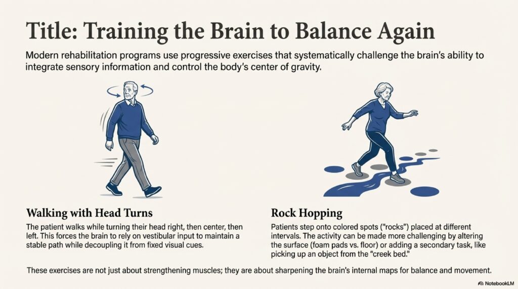
- Gaze stabilisation — VOR x1: Maintain fixation on a forward target during horizontal head oscillation (1 minute). Progress to lateral targets: turn head left, maintain fixation on left target (1 minute); repeat right. Complete front-left-right cycle five times.
- Neck flexion/extension: Maximum comfortable range flexion and extension while seated. Activates cervico-ocular reflex and desensitises cervicogenic dizziness contribution.
- Saccades and smooth pursuit: Horizontal and vertical eye movements to sequentially placed targets, 10 seconds each direction. Progress to 20 seconds as tolerance improves.
- Static balance — standing on firm surface: Erect stance, eyes open (1 minute), progress to eyes closed. Advance to next level when 5 minutes eyes-closed is achieved without upper limb support.
- Single-leg stance: 10 seconds each leg, alternating, five cycles. Progress to eyes closed. Clinician should supervise with arms out for safety.
- Ball tracking: Hold target object, move horizontally left-to-right while maintaining fixation (five repetitions). Repeat in vertical plane. Trains VOR x2 substitution strategy.
- Dynamic gait with gaze stabilisation: Walk while maintaining fixation on a fixed target or held object. Combines locomotor and vestibulo-ocular demands — approximates real-world functional requirements.
Uncompensated Unilateral Vestibular Hypofunction — Vestibular Rehabilitation Evidence-Based Protocol
This presentation is distinguished from acute vestibular neuritis by chronicity: symptoms persist beyond the expected natural compensation window (typically >3 months post-onset) without significant functional recovery. VOR gain deficit on vHIT and persistent caloric asymmetry confirm inadequate central compensation. These patients require a more intensive and structured programme than acute presentations.
Bed-Level Exercises
- Controlled breathing with sequential visual fixation (wall and ceiling targets, 30 seconds each position)
- Supine-to-sitting transition training with forward fixation
- Cat-cow stretch (quadrupedal): Inhale with spinal extension (hyperlordosis), exhale with spinal flexion (hyperkyphosis); coordinates breathing with trunk proprioceptive input
- Single knee-to-chest: Hold ipsilesional knee to chest, extend, repeat contralesionally
- Double knee-to-chest: Both knees held with hand assistance; passive lumbar flexion
- Hip rotations: Feet flat, knees flexed, rotate hips laterally keeping knees together; vestibular-independent proprioceptive training
- Weighted leg extensions: Pillow under affected leg, 2 kg ankle weight, extend leg — maintains lower limb strength during rehabilitation
Sitting Exercises
- Gaze stabilisation cycle: Forward fixation (1 minute), left lateral (1 minute), right lateral (1 minute); repeat cycle five times
- Neck flexion/extension through maximum comfortable range while seated
- Isometric neck strengthening: Hands clasped behind occiput, press forward while resisting with neck extensors; builds cervical proprioceptive contribution to postural stability
Standing Exercises — Firm Surface
- Static balance progression: Eyes open (1 minute), progressing to eyes closed (target 5 minutes) before advancing
- Single-leg stance: 10-second holds alternating, five cycles; progress to eyes closed
- Reading while walking: Walk while reading a held text — directly challenges VOR during self-generated locomotion
- Rhythmic lateral movement: Lateral trunk and arm movements for 3 minutes (increase to 5 minutes); dynamic postural challenge with rhythm cues
- Perturbation training: Clinician applies brief manual anterior and lateral perturbations (2-second pushes); develops reactive postural responses
- Ball tracking: Horizontal and vertical object tracking with visual fixation
- Target walking: Walk while maintaining continuous fixation on a fixed distal target
Foam Surface Exercises (Supervised)
Foam surface training removes stable plantar proprioceptive input, increasing reliance on residual vestibular and visual signals. Supervision is mandatory at this level. All foam exercises require a clinician or assistant positioned for fall protection.
- Erect standing on foam: 30 seconds eyes open, progressing to eyes closed (target 5 minutes eyes open before advancing)
- Foam dancing: Slow lateral hip and arm movements, 3 minutes (progress to 5 minutes)
- Foam marching: Military-style stepping on foam surface, then walking with lateral head turns
Bilateral Vestibulopathy — Protocol
Bilateral vestibulopathy (BV) presents a distinct challenge within the vestibular rehabilitation evidence-based protocol: substitution strategies rather than adaptation are the primary rehabilitation target, since the error signal driving cerebellar adaptation is absent or severely attenuated bilaterally. The Bárány Society diagnostic criteria (Strupp et al. 2017) define BV as bilateral caloric paresis (<6°/s combined) or bilateral reduced vHIT gain (<0.6 in the horizontal plane). These patients characteristically report oscillopsia during head movement and inability to walk in darkness.
Rehabilitation goals in BV are substitution-focused: maximising visual and somatosensory contributions to postural control. Exercise progression follows the same hierarchical sequence as UVH but with slower advancement criteria and indefinite maintenance programmes. Full compensation is rarely achieved; the clinical goal is functional independence with safety strategies for low-light and uneven surface environments.
- Bed: Breathing coordination, sequential visual fixation with wall and ceiling targets, supine-to-sitting transitions, cat-cow stretches
- Sitting: Gaze stabilisation cycle (front, left, right — five complete cycles), neck flexion/extension, isometric neck strengthening
- Standing on firm surface: Static balance progression, single-leg stance, rhythmic lateral movements (3–5 minutes), perturbation training, ball tracking (horizontal and vertical), target walking
- Foam surface (supervised): Erect standing 30 seconds progressing to 5 minutes, slow lateral movements (3–5 minutes), military marching on foam
Imbalance and Elderly Patients — Vestibular Rehabilitation Evidence-Based Protocol
Fall risk management is the primary clinical objective in presbyvestibulia and age-related balance decline. Neuroplasticity is preserved in healthy ageing but proceeds more slowly; exercise dosing should allow longer consolidation periods before progression. Comorbidities — osteoarthritis limiting range of motion, peripheral neuropathy reducing plantar proprioception, cardiovascular conditions limiting exercise tolerance — necessitate protocol individualisation. Mirror feedback is clinically useful in this population to augment visual postural cues.
Bed-Level Exercises
- Single knee-to-chest holds, each leg separately
- Double knee-to-chest with hand assistance
- Hip rotations: Flexed knees, rotating hips laterally with knees together
- Bridge with arm raises: Lift hips while extending arms overhead, return to neutral; strengthens posterior chain and challenges trunk stability
- Quadrupedal limb extensions: Right arm with left leg extended, alternating — diagonal pattern trains cross-limb coordination
- Cat-cow stretch with breathing coordination
- Sequential visual fixation with wall and ceiling targets
- Supine-to-sitting transition training with forward fixation
Sitting Exercises
- Gaze stabilisation cycle: Front, left, right — five complete cycles
- Neck flexion/extension through maximum comfortable range
- Floor-to-overhead reach: Bend forward from seated position, retrieve object from floor, raise to overhead (five repetitions); replicates functional ADL movement
- Isometric neck strengthening: Resisted forward press
Standing Exercises — Firm Surface
- Balance progression: Eyes open (1 minute), eyes closed (progress to 5 minutes)
- Single-leg stance: 10-second alternating holds, five cycles; progress to eyes closed
- Rhythmic large-movement training: Exaggerated lateral limb and trunk movements, 3–5 minutes — high-amplitude movement cues improve proprioceptive awareness
- Perturbation training: Clinician-applied anterior and lateral pushes (2 seconds)
- Floor-to-overhead reach in standing: Challenges balance during combined trunk flexion and upper limb elevation
- Ball tracking: Horizontal and vertical
- Large-step walking: Walk with exaggerated stride length while fixating on distal target; improves gait confidence and step clearance
Foam Surface Exercises (Supervised, in front of mirror)
- Erect standing on foam: 30 seconds eyes open, progressing to eyes closed (5 minutes target)
- Floor-to-overhead reach on foam: Challenges balance during movement
- Slow lateral movements on foam: 3–5 minutes
- Military marching on foam surface
Functional Mobility Training (ADL-Specific)
Clinically important functional movement patterns should be explicitly trained:
- Chair rise: Hands on chair arms, trunk forward lean, push through arms to standing. Pause in standing before initiating gait — allows vestibular-ocular stabilisation before locomotion.
- Floor object retrieval: Move object near stable support, use support surface to lower via knee bend, retrieve, use support to return to upright. This strategy reduces fall risk during a high-frequency ADL.
- Bed transfer: Log roll to lateral decubitus, lower legs over edge, use upper limb push to sitting. Pause in sitting with forward fixation before standing.
PPPD and Visual Vertigo — Protocol
Within the vestibular rehabilitation evidence-based protocol framework, Persistent Postural Perceptual Dizziness (PPPD) is defined by the Bárány Society diagnostic criteria (Staab et al. 2017) as chronic dizziness (>3 months) characterised by non-spinning dizziness or unsteadiness that worsens with upright posture, self-motion, and exposure to complex or moving visual environments. It frequently emerges following an acute vestibular event, with anxiety-driven hypervigilance perpetuating symptoms beyond physical recovery.
The fundamental pathophysiology involves mismatch between special proprioception (macular and cristal input) and general proprioception (muscular, ligamentous, and articular mechanoreceptors) during postural control, modulated by elevated autonomic arousal. Clinical reasoning: the adaptation pathway alone is insufficient for PPPD because the underlying dysfunction is cortical hypervigilance rather than peripheral VOR gain deficit. Combined VRT and cognitive-behavioural therapy (CBT) achieves 70–75% significant improvement rates (Staab et al. 2017). SSRIs (sertraline, fluoxetine) are appropriate pharmacological adjuncts in moderate-to-severe cases.
Key assessment tools for PPPD include: optokinetic test, Subjective Visual Vertical with moving visual background, and subjective midpoint assessment. These serve dual roles as diagnostic tools and as rehabilitative stimuli for visual motion desensitisation.
PPPD Exercise Protocol
A distinguishing feature of PPPD rehabilitation is the integration of cognitive loading during exercise. Recording video of first attempts and reviewing with the patient demonstrably reduces catastrophising and builds objective self-efficacy evidence.
- Sitting: Gaze stabilisation cycles (front, left, right), neck flexion/extension, floor-to-overhead ball reaches (five repetitions)
- Standing with concurrent mental task (dual-task protocol): Serial subtraction or word-list recall performed simultaneously with balance exercises. Static balance progression (eyes open/closed), single-leg stance cycles, rhythmic lateral movements (3–5 minutes), perturbation training, ball tracking (horizontal and vertical), target walking
- Foam surface with dual-task in front of large mirror: Erect standing 30 seconds progressing to 5 minutes, slow lateral movements (3–5 minutes), military marching, perturbation training. Mirror provides real-time visual postural feedback and simultaneously functions as an optic flow stimulus for graded visual desensitisation
- Graded environmental exposure: Structured hierarchy from low-stimulus environments (quiet room) toward high-stimulus environments (shopping centres, moving traffic) — graded exposure reduces conditioned avoidance responses
Contraindications and Precautions
Absolute contraindications to commencing VRT under this vestibular rehabilitation evidence-based protocol include: active central vestibular lesion requiring neurosurgical or neurological management (stroke, demyelinating episode, acoustic neuroma requiring re-intervention), unstable cardiovascular disease precluding exercise, and acute exacerbation of Menière’s disease with frequent endolymphatic hydrops attacks (>2 per week). In Menière’s, VRT is appropriate during stable inter-attack periods; active attack phases require medical stabilisation first.
Relative contraindications requiring protocol modification: cervicogenic dizziness as primary driver (add cervical physiotherapy), significant peripheral neuropathy (increase standing support requirements), high-dose vestibular suppressant use (taper under physician supervision before commencing VRT), severe anxiety or PTSD (initiate CBT concurrently), and significant orthopaedic limitations (modify exercise postures accordingly).
Halt exercises and reassess if the following occur: new severe headache during exercise, unilateral motor or sensory deficit, new diplopia, sudden hearing loss or significant increase in tinnitus, or symptoms not resolving within 30 minutes of ceasing exercise.
Inter-professional Referral Guidance
Applying this vestibular rehabilitation evidence-based protocol within a coordinated inter-professional framework optimises outcomes. VRT is most effective when delivered within a coordinated inter-professional framework. The following referral pathways should be considered:
- Refer to neurologist when: central vestibular pathology is suspected on examination (direction-changing nystagmus, gaze-evoked nystagmus, skew deviation, cerebellar signs), neuroimaging findings are abnormal, or VRT fails to produce expected gains at 8 weeks
- Refer to vestibular physiotherapist when: patient has bilateral vestibulopathy, complex comorbidities limiting home programme safety, failed home-based programme, or requires supervised foam and perturbation training
- Refer to audiologist when: sensorineural hearing loss accompanies vestibular symptoms (labyrinthitis, Menière’s, acoustic neuroma), VEMP interpretation is required, or BERA is indicated
- Refer to clinical psychologist/psychiatrist when: PHQ-9 or GAD-7 scores indicate moderate-to-severe depression or anxiety, PPPD diagnosis is established, or patient demonstrates significant avoidance behaviour limiting rehabilitation participation
- Refer to neuro-otologist or skull-base surgeon when: acoustic neuroma is identified on MRI, superior semicircular canal dehiscence is suspected, or perilymphatic fistula cannot be excluded
Key Clinical Principles of the Vestibular Rehabilitation Evidence-Based Protocol
- Progression criteria: Each exercise level has defined duration targets and stability criteria before advancement. Do not advance on a time-based schedule alone; advance on demonstrated performance criteria.
- Supervision requirements: Foam surface and perturbation exercises require clinician or trained assistant presence for fall protection.
- Optimal dosage: 3–5 sessions/day, 10–15 min/session, minimum 6 weeks (Level A evidence). Frequency of practice is the strongest neuroplastic driver; duration per session is less critical than repetition frequency.
- Vestibular suppressant interaction: Suppressants attenuate the adaptation error signal. Taper and cease vestibular suppressants prior to commencing adaptation-based VRT where clinically safe.
- Documentation: Record first attempts with video or standardised scoring for patient motivation and to demonstrate objective progress. Serial DHI and FGA scores provide medicolegal and clinical quality data.
- Individualisation: Tailor protocols to the specific vestibular diagnosis, side of lesion, symptom profile, comorbidities, and patient functional goals.
References
- Hillier SL, McDonnell M. Vestibular rehabilitation for unilateral peripheral vestibular dysfunction. Cochrane Database of Systematic Reviews. 2011;(2):CD005397. [PubMed]
- McDonnell MN, Hillier SL. Vestibular rehabilitation for unilateral peripheral vestibular dysfunction. Cochrane Database of Systematic Reviews. 2015;(1):CD005397.
- Whitney SL, Sparto PJ. Principles of vestibular physical therapy rehabilitation. NeuroRehabilitation. 2011;29(2):157–166.
- Staab JP, Eckhardt-Henn A, Horii A, et al. Diagnostic criteria for persistent postural-perceptual dizziness (PPPD): Consensus document of the Committee for the Classification of Vestibular Disorders of the Bárány Society. Journal of Vestibular Research. 2017;27(4):191–208.
- Strupp M, Kim JS, Murofushi T, et al. Bilateral vestibulopathy: Diagnostic criteria Consensus document of the Classification Committee of the Bárány Society. Journal of Vestibular Research. 2017;27(4):177–189.
- Lacour M, Tighilet B. Plastic events in the vestibular nuclei during vestibular compensation: The brain orchestration of a “deafferentation” syndrome. Restorative Neurology and Neuroscience. 2010;28(1):19–35.
- Deveze A, Bernard-Demanze L, Xavier F, Lavieille JP, Elziere M. Vestibular compensation and vestibular rehabilitation. Current concepts and new trends. Neurophysiologie Clinique. 2014;44(1):49–57.
- Hall CD, Herdman SJ, Whitney SL, et al. Vestibular rehabilitation for peripheral vestibular hypofunction: An evidence-based clinical practice guideline. Journal of Neurologic Physical Therapy. 2016;40(2):124–155.
- Herdman SJ. Vestibular rehabilitation. Current Opinion in Neurology. 2013;26(1):96–101.
- Bhattacharyya N, Gubbels SP, Schwartz SR, et al. Clinical practice guideline: Benign paroxysmal positional vertigo (update). Otolaryngology–Head and Neck Surgery. 2017;156(3_suppl):S1–S47.
Medical Disclaimer: This article is a clinical reference for qualified healthcare professionals. It is not a substitute for individual clinical judgment, local guidelines, or direct patient assessment. Treatment should be personalised based on specific diagnosis, examination findings, comorbidities, and patient goals.